
Nessun prodotto nel carrello.
6-12 Months, Augma Bone Cement Academy, Bond Apatite®, Bone Cement, Bone Cement Expert, Clinical Cases, Clinical Indication, Clinician, Cyst Enucleation, Dental Notation, Images, Lower Left Incisor, Lower Right Incisor, Media, Post-Op Period
Large Central Ossifying Fibroma of the Anterior Mandible
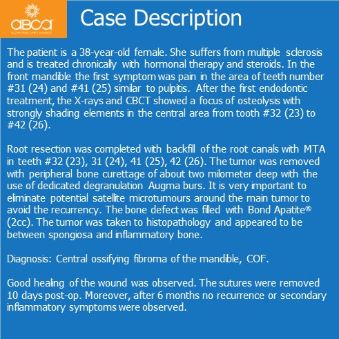
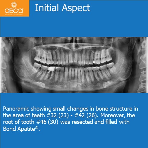
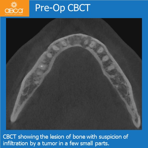
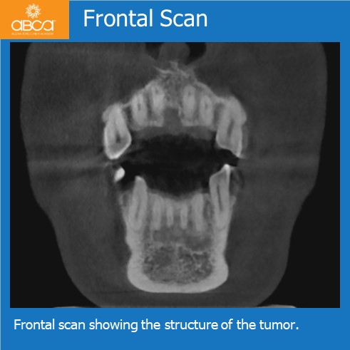
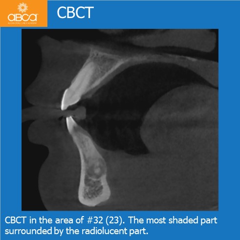
The patient is a 38-year-old female. She suffers from multiple sclerosis and is treated chronically with hormonal therapy and steroids. In the front mandible the first symptom was pain in the area of teeth number #31 (24) and #41 (25) similar to pulpitis. After the first endodontic treatment, the X-rays and CBCT showed a focus of osteolysis with strongly shading elements in the central area from tooth #32 (23) to #42 (26).
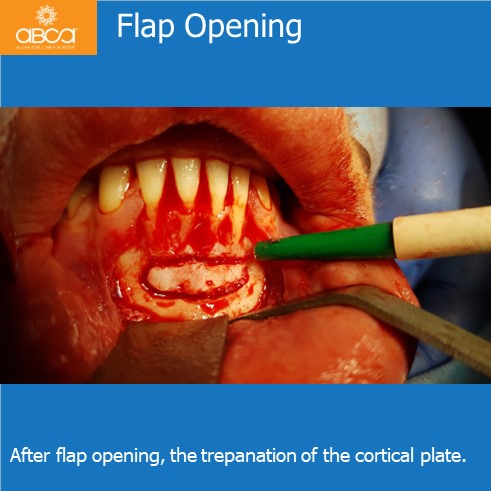
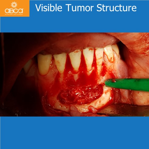
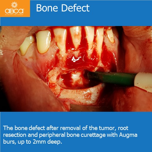
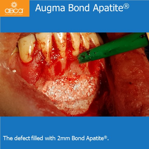
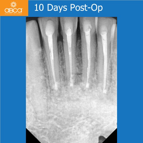
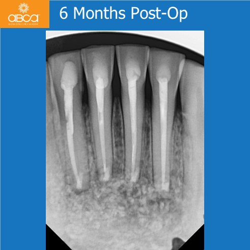
Root resection was completed with backfill of the root canals with MTA in teeth #32 (23), 31 (24), 41 (25), 42 (26). The tumor was removed with peripheral bone curettage of about two milometer deep with the use of dedicated degranulation Augma burs. It is very important to eliminate potential satellite microtumours around the main tumor to avoid the recurrency. The bone defect was filled with Bond Apatite® (2cc). The tumor was taken to histopathology and appeared to be between spongiosa and inflammatory bone.
Diagnosis: Central ossifying fibroma of the mandible, COF.
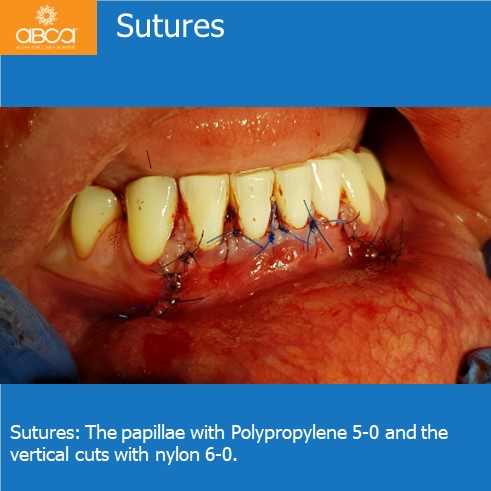
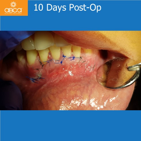
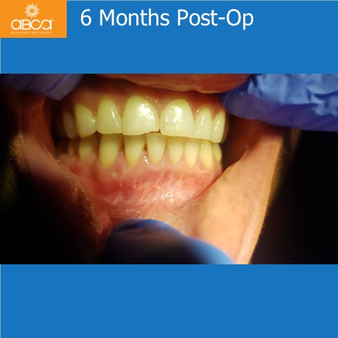
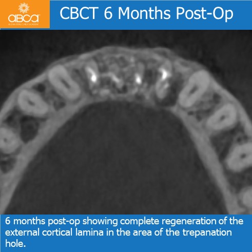
Good healing of the wound was observed. The sutures were removed 10 days post-op. Moreover, after 6 months no recurrence or secondary inflammatory symptoms were observed.